We are living through a technological revolution. The big tech companies are working to make our interactions with machines more intuitive, aiming to create the illusion that we are communicating with a person. Our digital interactions often involve a series of taps, spoken words, and specific hand gestures to engage with devices or AI technologies in our homes, workplaces, in virtual environments (VR), and augmented reality (AR). As these advancements become a reality, the critical question arises: how we can train these technologies to read between the lines and understand us as humans do? This isn’t just about convenience. It’s about significantly enhancing our sense of agency, the intuitive feeling of control and the connection we experience with the tools we use. The future of Human-Machine Interaction will rely not only on our direct instructions but also on the silent and instinctive signals conveyed through our bodies and facial expressions.
Why Facial Signals Matter While gestures and voice commands are an essential to Human-Machine Interfaces (HMI), they often miss the valuable layer of communication offered by facial expressions. Our faces continuously emit electrophysiological signals, subtle muscle activations that reveal our emotions and intentions, many of which occur involuntarily. Traditional cameras struggle to capture these subtle signals due to environmental challenges such as lighting and positioning. Facial Electromyography (fEMG) offers a solution by measuring the electrical activity of facial muscles. It can detect even the smallest, often unconscious expressions that are not visible to the naked eye. By combining high sensitivity fEMG monitoring with machine learning (ML) models, and incorporating a personal calibration process, we can develop interfaces that truly understand and adapt to the user’s emotional state.
The Future of Adaptive Interfaces Imagine you’re in the middle of cooking a complex dish, with both hands busy and your attention focused on the ingredients. Step-by-step instructions from your AI assistant guide you through the recipe. But as the pace and complexity increase, what if the system could detect subtle signs of strain? Not just from you asking it to repeat instructions or scrolling back, but from almost imperceptible cues like a furrowed brow, raised eyebrows, or a brief pause, which indicate that you’re feeling confused or overwhelmed. By integrating fEMG into the AI technologies, the system gains extraordinary insights. It can detect these early, often invisible cues that signal a user nearing his cognitive limit. Picture the system intelligently simplifying the visual display, slowing the pace of instructions, or even gently suggesting a brief, well-timed pause. This isn’t just a response to actions, it’s a real-time adjustment to the user’s internal, non-conscious state. Such an adaptive interface doesn’t just guide, it supports and optimizes human performance by understanding the subtle, silent language of the body.
Looking Ahead Physiological signals, particularly fEMG, expand the possibilities for human-machine interaction. They reveal moments of focus, hesitation, or emotional responses that might otherwise go unnoticed. This makes interfaces more adaptive, intuitive, and genuinely aware of human emotions and intentions. We are just at the beginning of this journey. As these technologies continue to evolve, we will witness machines that not only respond to our actions but also react to our feelings and intentions in real-time. This significant progress represents a step toward creating interfaces that feel less like tools and more like collaborators in our daily lives.
When developing wearable sensor patches capable of providing medical-grade monitoring outside of clinical settings, we must begin by appreciating that creating a fully functional system involves some essential building blocks, all of which must work together. From the technological perspective, developing the patch may appear to be the first step. However, being able to design a working patch in a way that is compliant with specific guidelines and will be accepted by regulatory authorities must be learned and applied.
Developing a multimodal solution for at-home use entails significant effort and expertise. Central to this is understanding the problem you are trying to solve, especially when developing electrode patches intended for a diverse range of applications, from sleep monitoring and analysis to sports rehabilitation. The level of knowledge and expertise required for each application can be immense. Sleep alone is a huge field, with over 40 different sleep-related disorders, each of which must be fully understood. This entails understanding the medical manifestation of the particular disorder, the different technologies involved, and how existing technology is already being applied. This is before we even begin to address the various commercial issues and market needs.
1. A Multimodal Solution Requires A Multifaceted Team
Central to an endeavor focused on developing a multimodal wearable solution is building a multifaceted team comprising all the necessary expertise prior to initiating development. Such a team cannot be siloed, as there are many key areas where team members must fully understand the intricacies of different domains. For example, in the initial stages of designing electrode patches, those responsible for developing the electronic components must also have a deep understanding of the fundamental issues relating to the different interfaces the patches will interact with — in our case, primarily the skin and neural interfaces. Similarly, data scientists working on the algorithms must also appreciate the limitations of the hardware, as well as the market requirements and specific medical domain being addressed.
It is therefore incumbent on team leaders to draw on talented, multidisciplinary professionals when developing a new solution. Encouraging dexterous team members able to immerse themselves in the various domains related to wearable technology will prove a key element in successfully introducing a medical-grade wearable technology to market.
2. Collaboration With Industry Players Is Key
Developing a medical-grade wearable solution cannot occur in a vacuum, especially when it comes to the relatively small ecosystem involved in the application of flexible electronics for electrophysiological monitoring. From the outset, it is essential to tap into the wider industry for insights, collaborating with other players, such as leading researchers and sleep centers, who can provide essential feedback based on their own experiences.
This approach can entail certain frustrations, especially in the earliest R&D stages, which can result in initial angles being rejected outright by a traditionally risk averse industry. However, such feedback will ultimately prove invaluable, not just in determining which direction to take but also understanding the factors that may cause a solution to be rejected; in many instances, this will come down to cost or lengthy production times. Thus, sourcing knowledge from the wider industry can encourage further creativity and, through perseverance and continually trialing different concepts and approaches, result in the ability to develop a solution that fully addresses the needs of the market.
3. Ensure Ease Of Use
Addressing usability and simple self-application is central to developing an at-home solution, ensuring that the final product does not require intensive training while also providing clinical-grade results. This boils down to the design — how the device is physically designed and how much room for error can be allowed when operating it. This must be addressed from the earliest stages of development, taking into account the users — be they physicians or patients — and working out all the various user interface issues. The way in which a company designs a medical wearable technology will directly correlate to the frequency of errors. Therefore, tackling design interface issues at the earliest stages will pave the way for developing a reliable and robust device suitable for medical applications.
One particular challenge arises when developing a method to affix the electro-patches to the body in a way that ensures highly accurate readings, in different environments and for extended periods of time, without impacting users comfort. Paramount to developing a viable wearable solution is ensuring that the system has a small form factor. In the context of wearable electrodes, developers must ensure that the patch attaches to the skin seamlessly and is transparent so that users can perform naturally without having their lives impacted by the system. This will naturally relate to the weight and dimensions of the electronic system. In the case of X-trodes, this required collaborating with leading companies in the fields of printing and adhesives. Tapping into their expertise enabled the development of a dry-printed electro-patch that is easily self-administered by patients, does not require any gel or adhesive, and is flexible enough to comfortably conform to the natural contours of the body, enabling continuous monitoring in any environment.
Addressing usability and simple self-application is central to developing an at-home solution, ensuring that the final product does not require intensive training while also providing clinical-grade results. This boils down to the design — how the device is physically designed and how much room for error can be allowed when operating it.
dry-printed electro-patch that is easily self-administered by patients
4. Ensure Efficacy Of Data
The ultimate goal when developing a wearable monitoring solution is to provide the user, be they end users or medical practitioners, with actionable insights. However, this may not necessarily be the data that the system provides directly. For example, while cardiac measurements can be provided by an ECG trace, a trained professional is still needed to analyze the data and provide a medical evaluation. In many aspects of medical practice, data is provided in a very crude form. However, the end goal is to have analyzed data that can provide actionable insights and medical advice. Achieving this in a stable manner requires a significant effort, made even more challenging when we try to shift to fully automated data analysis.
Overcoming this challenge requires a large amount of data preparation and signal processing routines, which must be adapted to provide data that can be analyzed and the bottom line provided automatically. Medical data is generally collected under relatively sterile conditions in controlled environments. An at-home solution must be able to provide that same quality of medical-grade data collected in real-life scenarios, outside of clinical settings. This requires removing as many external interferences as possible. When the end user is moving and performing natural activities, ensure that daily — or even high-impact — activity does not interfere with the system’s recording and processing capabilities. Thus, a properly designed system must address the dual challenge of ensuring that the device does not interfere with the user’s natural behavior while, at the same time, a person’s normal behavior does not impact the quality of data collection. This will ensure that, from the beginning, the data collected is as clean as possible, as data contaminated with lots of external artifacts will make the analysis extremely challenging.
Thus, a significant effort has to be put into developing algorithms, either by optimizing existing algorithms or developing entirely new ones, that can identify a variety of different conditions and allow the automation of processes that enable us to understand the data received — whether clean or contaminated — which can then be analyzed. A number of new approaches, such as neural network models and deep learning, are proving invaluable in their ability to make automated decisions without requiring a person to physically analyze and evaluate the data.
Conclusion
Designing and developing a medical-grade wearable solution, therefore, entails multiple interlocking components to ensure market acceptance and adoption by clinicians and end users. Ultimately, it boils down to the ability to provide insightful and actionable data through a user-friendly device in diverse environments. Wearable monitoring devices generate vast amounts of data essential for establishing the medical validity of the solution. Any glitch in the system can result in a loss of data, without which we cannot establish medical validity. Therefore, the ability to generate clean and validated data via a robust and usable system is integral in order to progress from proof of concept to development and eventual commercialization.
Facial expressions are a window into human emotions, offering valuable insights across psychology, neurology, and Human-Machine Interface (HMI) applications. From decoding micro-expressions for lie detection to monitoring emotional responses to stimuli like ads or music, facial expression analysis is a powerful tool. Facial expression analysis also plays a critical role in many clinical settings, aiding in the management of neurologic conditions like Parkinson’s disease, guiding Botox injections, and supporting rehabilitation after facial nerve palsy. In psychiatry, it provides objective measures for tracking affective states, while in HMI, it enhances neuroadaptive systems by gauging operator attention, satisfaction, or frustration. AI-driven emotional support systems can leverage facial expression analysis as a real-time feedback mechanism.
Traditional video-based facial expression analysis requires subjects to remain stationary in front of a camera and is highly sensitive to lighting conditions, making it impractical for use in dynamic environments or low-light settings. Existing electromyography (EMG) solutions rely on wired setups and bulky gel electrodes that hinder natural facial movements and limit mobility.
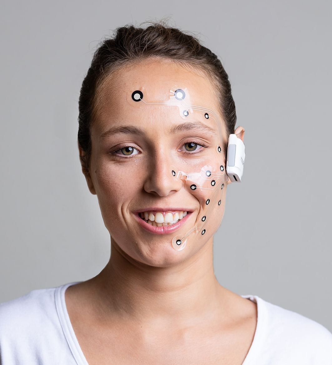
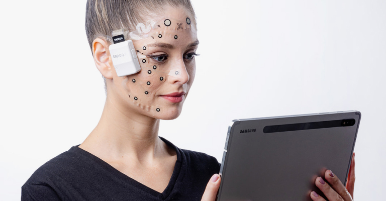
Enter X-trodes, a game-changer in facial EMG (fEMG) technology. X-trodes introduces thin, soft, flexible patches that collect fEMG signals non-invasively, enabling natural facial expressions and unrestricted movement. These patches are designed for real-world use, resistant to humidity, dust, and sweat, and seamlessly integrating with hats, glasses, or professional gear like helmets. With a sampling rate of up to 4KHz, X-trodes can capture even the briefest micro-expressions. The wireless design and advanced filtering make it the world’s first solution for fEMG collection on the move. In their latest work, Facial muscle mapping and expression prediction using a conformal surface-electromyography platform, Hila Man and colleagues demonstrate for the first time successful reconstruction of free facial expression from fEMG recordings and provide an anatomic framework for facial muscle activity analysis. A prior publication by Paul Funk and colleagues, Wireless high-resolution surface facial electromyography mask for discrimination of standardized facial expressions in healthy adults, demonstrated successful reconstruction of standardized expressions.
The versatility of X-trodes extends beyond fEMG. These patches can simultaneously collect EEG, EOG, ECG, and hand EMG, providing a comprehensive view of emotional and physiological states through brain activity, eye movements, blink rate, heart rate variability, and gestures. All modalities are FDA approved. This multi-modal capability makes X-trodes ideal for mental state studies.
X-trodes also supports simultaneous synchronized recordings of multiple subjects, useful in social interaction studies.
X-trodes’ wireless, high-resolution, and user-friendly design empowers researchers, clinicians, and engineers to unlock new possibilities in emotional and neurological analysis.
Here are some examples of publications utilizing this technology:
In the world of EEG research, a paradigm shift is underway. The conventional wisdom of prioritizing low impedance in pursuit of high-quality electrophysiological recordings is being reevaluated. Its time to redirect our attention toward a more meaningful metric: the signal-to-noise ratio (SNR).
For decades, the pursuit of low skin-electrode impedance held great importance. This belief stemmed from an era when amplifiers drew significant input currents and data points were meticulously plotted on graph paper. Back then, obtaining a quality signal required the use of silver-silver chloride electrodes, electrolytes, and even skin abrasion. The gold standard for EEG recording was set at an impedance of less than 5KΩ.
When coupled with sophisticated signal processing techniques researchers can now attain Impeccable recordings even when dealing with low amplitude signals, such as EEG.
Recordings with low amplitude signals
Fast forward to today, and modern amplifiers have revolutionized the landscape. These advanced devices boast high input impedance, rendering the concern for significant current draw obsolete. This breakthrough enables stable recordings to be achieved with substantially higher skin-electrode impedance values. When coupled with sophisticated signal processing techniques, researchers can now attain impeccable recordings even when dealing with low amplitude signals, such as EEG. This progress is particularly evident with the advent of cutting-edge technologies like dry printed electrodes.
As we embark on this new frontier, its imperative to reassess our assumptions and embrace the latest advancements in EEG recording. Rather than fixating on the impedance metrics of the past, our focus should shift towards optimizing the signal- to-noise ratio. By doing so, we open up doors to unprecedented insights and discoveries.
EEG
HIGH QUALITY RECORDING
RESEARCH
SIGNAL-TO-NOISE
SNR
For those eager to delve deeper into this transformation, there are several compelling studies in the literature that shed light on the subject: